
Portfolio
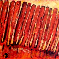
Leucophores in White

Cuttlefish skin camouflage system
The source image for this piece is a Scanning Electron Micrograph taken at Marine Biological Lab by Alan Kuzirian of cuttlefish skin.
Read MoreLeucophoresphores in Red 2

Cuttlefish camouflash subcellular structures based on Scanning Electron Micrograph (SEM) taken at Marine Biological Lab by Alan Kuzirian of cuttlefish skin.
Read More


Pneumotaxic Burst
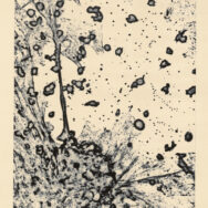
This artwork was made as part of Imperial College of London’s, Beautiful Science project, in collaboration wit Eric Dubuis of the Respiratory Pharmacology department. It is an etching print of neurons in the nodose ganglion of the vagus nerve trunk, strongly expressing Transient Receptor Potential (TRP) type channels which have been implicated in airway inflammatory response and the cough reflex.
The airways are innervated by nerves whose cell bodies originate in the nodose and jugular vagal sensory ganglia, which are located at the level of the first cervical vertebra. Activation of these cells by environmental irritants and inflammatory mediators in the lungs leads to central reflexes like cough. TRP channels are a new promising target for the development of drugs to treat cough and inflammatory lung diseases like asthma and emphysema. On the lower left side of the image, a convex reticulated structure depicts a vagal sensory neuron’s body; axons extend in an explosive pattern. The globular structures are transverse sections of dendrites and the smaller points are an abstraction of neurochemicals released during stimulation.
Read MoreSomata Fragments
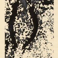
In Somata Fragments, also part of Imperial College of London’s Beautiful Science exhibition, we see a neuron’s body in a process of fragmentation which reflects the myriad stimuli, inputs and outputs that neurons have the capacity of transmitting and analyzing to create our complex bodily reality.
Read More